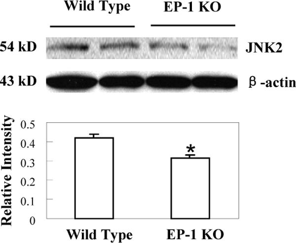
Figure 10. Decreased expression of JNK2 in EP1 knockout mice treated with LPS/D-GalN.

EP1 knockout mice and matched wild type mice were sacrificed 4 hours after LPS and D-GalN injection. The liver tissues were homogenized and the extracted proteins were subjected to SDS-PAGE and Western blot analysis to determine the protein level of JNK2. Western blot for β-actin was used as the loading control. Reduced JNK2 protein was observed in the EP1 knockout livers when compared to the wild type controls. The lower panel represents the ratio between JNK2 and β-actin by densitometry analysis (* p < 0.01 compared to wild type mice).
