
Figure 6. Increased expression of JNK2 in COX-2 transgenic mice.
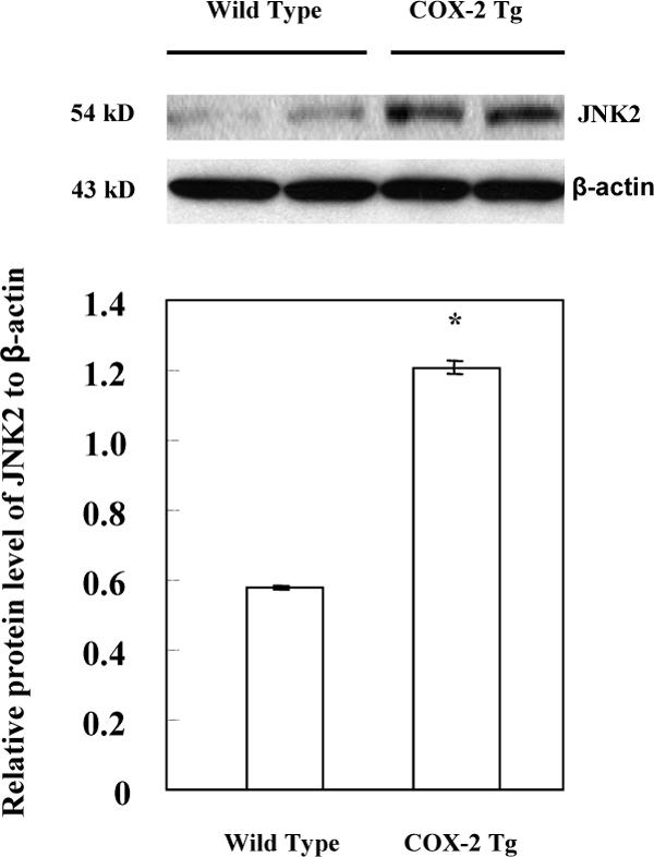
The liver tissues from the COX-2 transgenic mice and their matched wild type mice were homogenized. The cellular proteins were subjected to SDS-PAGE and Western blot analysis to determine the protein level of JNK2. Western blot for β-actin was used as the loading control. Higher level of JNK2 was observed in the liver tissues from the COX-2 transgenic mice when compared to the wild type mice. The lower panel represents the ratio between JNK2 and β-actin by densitometry analysis (* p < 0.01).