
Figure 7. Increased phosphorylation of JNK in COX-2 transgenic mice treated with LPS/D-GalN.
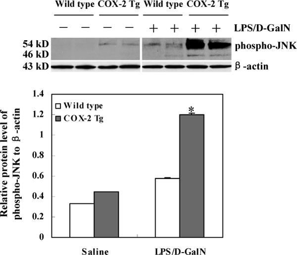
The COX-2 transgenic mice and matched wild type mice were sacrificed 4 hours after LPS/D-GalN injection. The liver tissues were homogenized and the extracted proteins were subjected to SDS-PAGE and Western blot analysis using the antibody against phospho-JNK (Cell Signaling Technology, Danvers, MA). Western blot for β-actin was used as the loading control. The lower panel represents the ratio between phosphorylated p54-JNK2 and β-actin by densitometry analysis. *ρ<0.01 compared to wild type mice treated LPS/D-GalN or COX-2 Tg mice treated with saline.