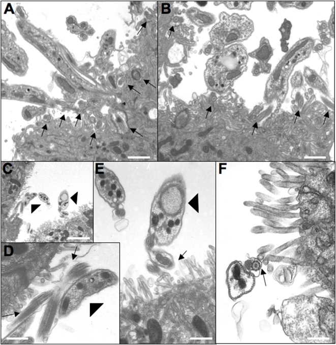
Figure 7. Representative images from sections of salivary glands.
(A, B) Salivary glands isolated from flies infected with wild type AnTat1.1. Note that the flagella (marked with arrows) are in very close contact with the epithelial cells and often appear completely entangled with surface microvilli of the salivary gland. (C–F) Salivary glands from flies infected with Δprocyclin. (D) and (E) are higher magnification views of those areas in C marked by arrowheads. Arrows mark the flagella adhering to the epithelia. Note the much lower density of parasites, and the much less pronounced interaction of Δprocyclin with the epithelium compared to wild type AnTat1.1. Bars in (A) = 1.1 µm; (B) = 0.8 µm; (C) = 2.4 µm; (D) = 0.6 µm; (E) = 0.6 µm; (F) = 0.44 µm.

