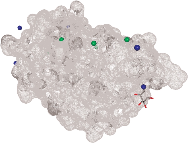
Figure 10. The structure of Anguilla anguilla agglutinin protein (PDB entry 1K12).
The binding sites predicted by Ligsite CSC are colored in green and the binding sites predicted by the pattern-based method are colored in blue. The protein surface is rendered in gray and the ligand is in the stick form. The LigsiteCSC predictions are over 10Å away from any atom of the ligand, while one of pattern-based predictions is 0.67Å away from one of the ligand's atoms. Only 4 predictions by Ligsite CSC and by the pattern-based method are visible; the remaining predictions are on the other side of the protein.

