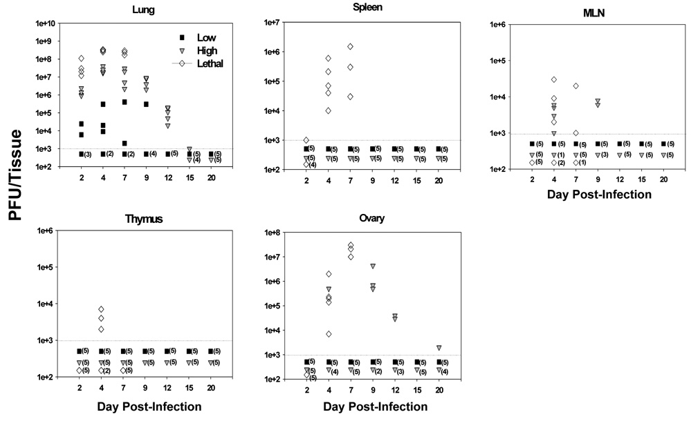
Figure 5. VACV-Ova was not detectable in the spleen following high dose infection.
C57BL/6 mice were infected i.n. with the low (black squares), high (gray triangles), or lethal (open diamonds) dose of VACV-Ova. Lung, spleen, MLN, thymus, and ovary were harvested at the indicated timepoints and VACV-Ova titers from each whole tissue determined. Each symbol represents one mouse, with numbers in parentheses indicating the number of mice with organ titers below the limit of detection (103 PFU).

