
Figure 9.
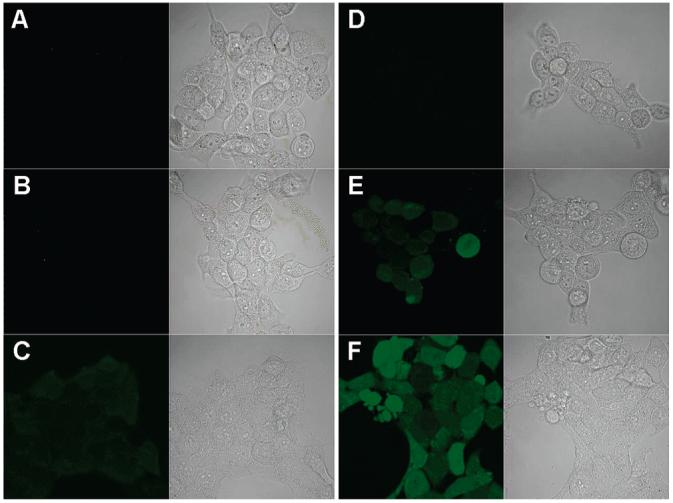
Confocal images of HaCaT cells stained with 5 μM PF1; ex/em, 488/505-550 nm. (A) Cells were kept in the dark and incubated with 5 μM PF1 for 5 min at RT; left, confocal fluorescence microscopy; right, brightfield transmission microscopy. (B) Same as part A except that cells were incubated with 0.5 μM (γ-CyD)2/C60 for 2 h at 37 °C before staining with PF1. (C) Same as part B except that cells were incubated with 30 μM C60(OH)24. (D-F) Same as parts A-C, respectively, except that cells were then exposed to UVA (15 J/cm2) for 10 min.