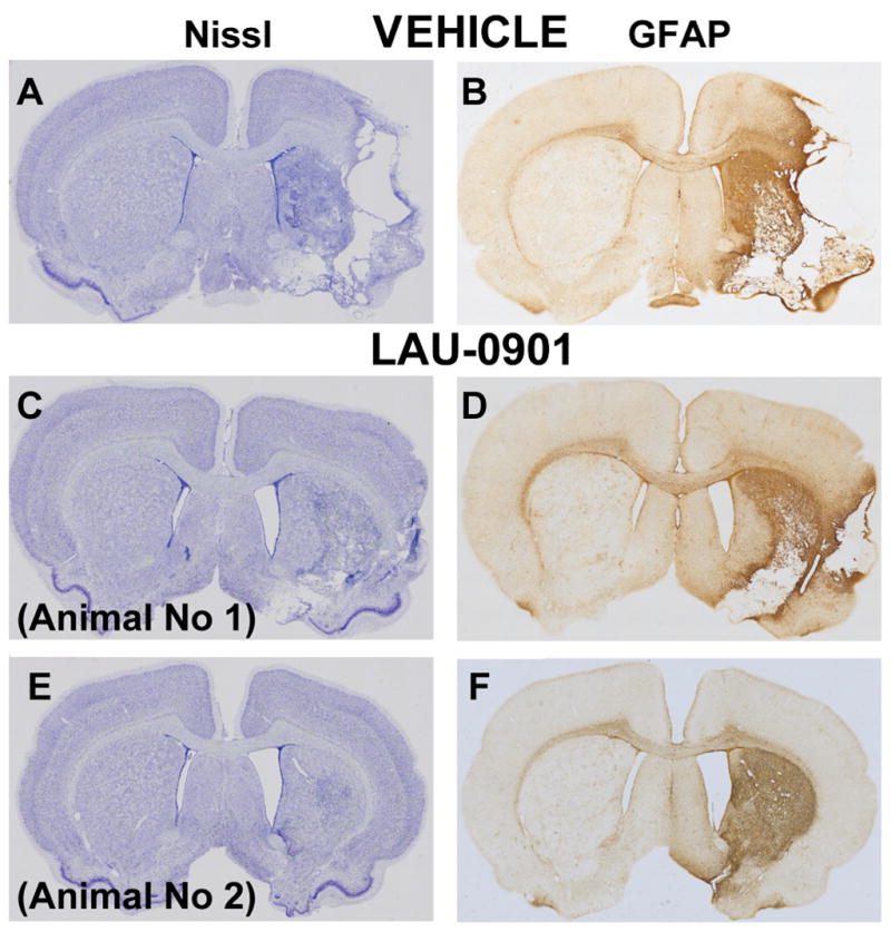
Figure 4.

Computer generated MosaiX processed images (Carl Zeiss MicroImaging, Inc, Thornwood, NY) of Nissl (Panels A, C and E) and GFAP (Panels B, D and F) paraffin-embedded brain sections at coronal level (bregma +1.2 mm) from a rat treated with vehicle (Panels A and B) and two rats treated with LAU-0901 (Panels C–F). The saline-treated rat shows typical appearance of cystic necrosis, and pan-necrosis involves the entire neocortical thickness, extending to subjacent regions (Panels A–B). In contrast, the two rats treated with LAU-0901 show less extensive damage (Panels C–F).
