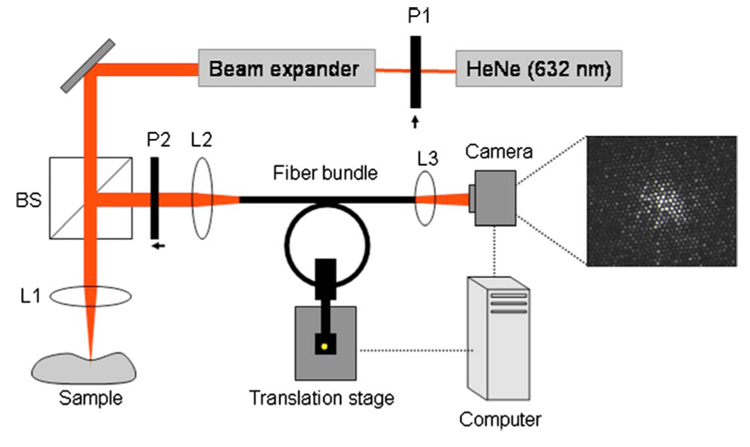
Fig. 1.
Experimental setup for LSI. To obtain laser speckle images through the fiber bundles, light (632 nm) from the helium-neon source was focused on a sample, and the resulting speckle pattern was imaged on the distal end of the fiber bundle. The fiber bundle was inserted through plastic tubing wound over a 2-cm radius of curvature to mimic the human LAD curvature. The proximal end of the bundle was imaged using a CCD camera, and speckle images were obtained at a high frame rate. The fiber bundle was connected to a motorized stage that modulated the motion of the bundle (in the direction perpendicular to the page) to mimic human coronary motion over the cardiac cycle (L1, L2, L3=lenses; P1, P2=polarizers; BS =beamsplitter).

