
Fig. 4.
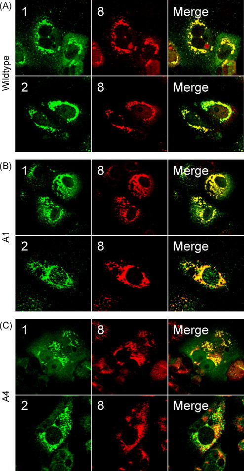
Immunofluorescence of SARS-CoV-infected cells. Vero cells on glass coverslips were infected with wildtype (wt, A), A1 (B), or A4 (C) viruses. Mutant designations are as outlined in Fig. 3. Cells were fixed and permeablized with methanol at 12 h p.i. Fixed coverslips were then stained for nsps 1 or 2 along with nsp 8 as a marker for replication complexes. Images were obtained using a Zeiss LSM 510 confocal microscope and were processed using Adobe Photoshop CS2 (9.0.2).