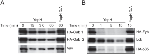
Figure 2. YopH dephosphorylation assay of several proteins.
A, Dephosphorylation assay for HA-Gab1, HA-Gab2 and Vav at different time-points, using 1 µg of GST-YopH or GST-YopH D356A. The assay was stopped by addition of sample buffer, and after SDS-PAGE, samples were transferred to nitrocellulose and tyrosine phosphorylation was detected by Western blot with anti-phosphotyrosine antibody. B, Dephosphorylation assay for HA-Fyb, Lck and HA-p85 was carried out as in A, but using shorter incubation times. Proteins used as substrates in these assays were obtained from HEK293 transfected with the corresponding plasmids and treated with pervanadate. Proteins were immunoprecipitated and distributed equally in different tubes for the several time-points of the assay.

