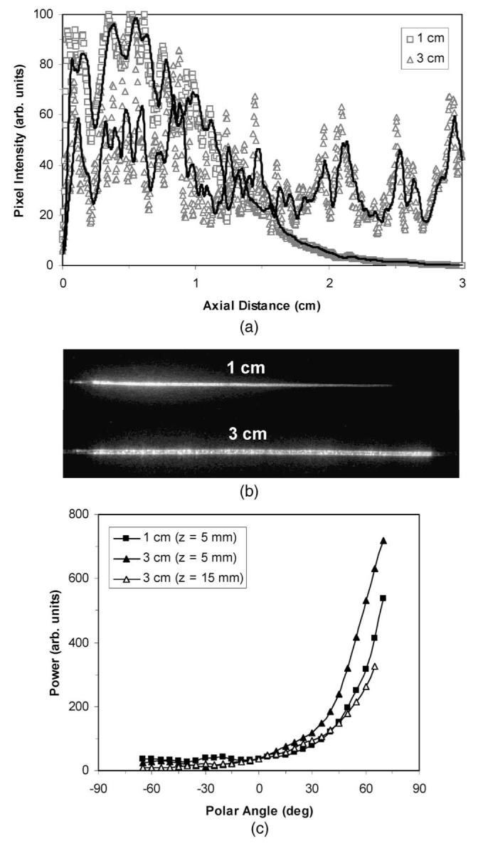
Fig. 6.

(a) Axial emission profiles and (b) images of 300-μm-diam diffusers created using nozzle-to-sample distances D of 1 and 3 cm. In (a), the curves were normalized based on the amount of light emitted over the entire 3-cm length measured using the integrating sphere system. The diffusion efficiencies over axial distances of 1, 2, and 3 cm were 82, 99, and 100%, respectively, for D=1 cm and 37, 68, and 91%, respectively, for D=3 cm. (c) Polar emission profiles of the diffusers in (a) and (b) at axial distances of 0.5 cm (D=1 and 3 cm) and 1.5 cm (D=3 cm only). In (c), the curves were normalized to have equal power at a polar angle of 0 deg (perpendicular to diffuser axis). In (a) to (c), the nozzle translation speed and blasting pressure were 3 mm/s and 40 psi, respectively.
