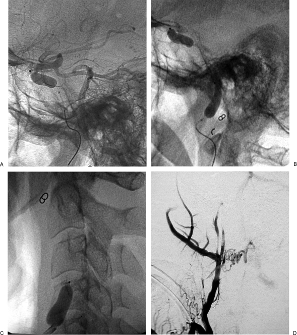
Figure 3.
Same case as Figs. 1 and 2. (A) Left vertebral artery injection was administered after placement of detachable balloon in cavernous segment of internal carotid artery (ICA). (B) Second balloon was inserted in petrous and distal cervical segment of ICA after detachment of the balloon in cavernous segment of ICA. (C) A third balloon that was positioned and detached in proximal cervical segment of ICA is seen. (D) Injection of right common carotid artery, in laterolateral view, shows complete devascularization of the tumoral mass after balloon occlusion of ICA and embolization of external carotid artery branches.

