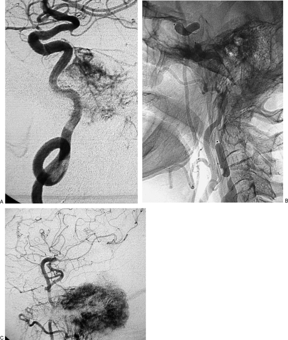
Figure 6.
Same case as Figs. 4 and 5. (A) Left internal carotid artery (ICA) injection, lateral view, shows tumoral vascular supply coming from the meningohypophyseal trunk and stenosis of the petrosal horizontal segment; coiling of midcervical portion of ICA, which precludes insertion of a stent, is also evident. (B) Left common carotid artery injection, lateral view cervical level, shows three detachable balloons in place and patency of the bypass. (C) Left common carotid artery injection, lateral view at cranial level, shows patency of bypass, intracranial anastomosis with middle cerebral artery, revascularization of supraclinoid portion of left ICA and tumoral blush coming from branches of external carotid artery.

