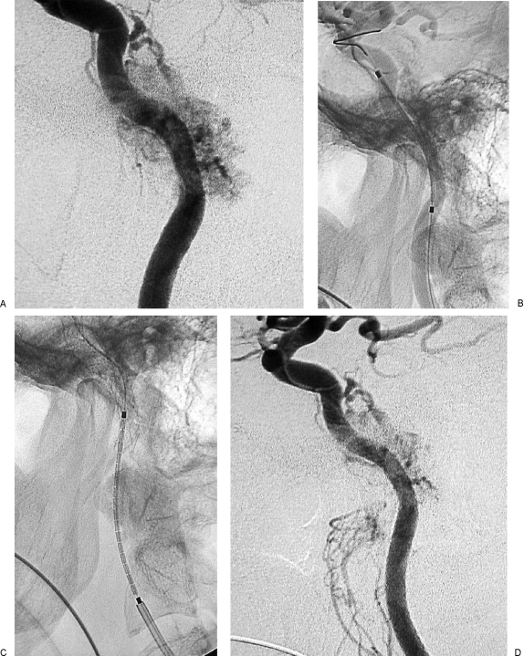
Figure 8.
Same case as Fig. 7. (A) Left internal carotid artery (ICA) injection, lateral view, shows tumoral supply from meningohypophyseal trunk and stenosis of distal cervical and petrosal vertical segments of ICA. (B) Left ICA, lateral view, shows insertion of Xpert stent in petrous segment of ICA. (C) Left ICA, lateral view, shows insertion of Xpert stent in the distal cervical segment, with 10 mm overlapping with the first stent. (D) Left ICA injection, lateral view, after insertion of two Xpert stents, shows disappearance of the previously seen stenosis and slight reduction of the tumoral blush.

