
Figure 5.
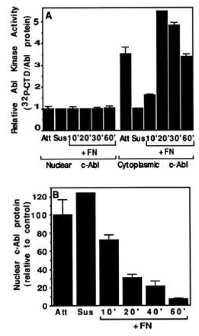
c-Abl activity and localization in quiescent cells. The 10T½ cells starved in 0.1% serum for 48 hr were trypsinized and then plated on FN in medium containing 0.1% serum. At the indicated times cells were harvested and fractionated. (A) Activity. Cells were either stably attached (Att) or trypsinized (Susp) and plated onto FN-coated plates. At the indicated times, cells were harvested and fractionated, and the c-Abl was immunoprecipitated from the lysates and the kinase activity was determined. The mean and standard deviation of three experiments are shown. (B) Nuclear c-Abl level. The relative levels of nuclear c-Abl at each time point after adhesion to FN were determined as in Fig. 2.