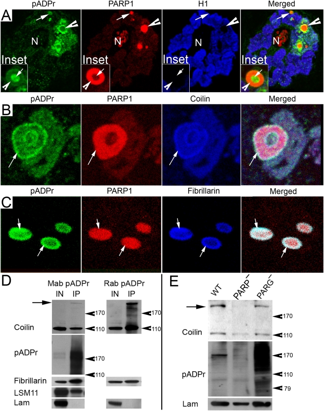
Figure 6. The components of Cajal body are targets for pADPr.
(A) The immunostaining of thick sections prepared from Parg27.1 mutant salivary gland is shown. “Free” nucleoplasmic bodies (arrow) and “chromatin-embedded” CBs (arrowhead) are shown. Specific antibodies were used: rabbit anti-pADPr (green) and mouse anti-histone H1 (blue). PARP1-DsRed (red) was visualized by DsRed autofluorescence. Inset. The single CB-like particle is magnified. Arrow indicates accumulation of pADPr in the CB cavity. Arrowhead shows enrichment of PARP1 protein in the CB matrix. (B, C) Immunostaining of Parg27.1 mutant salivary gland is shown. Specific antibodies were used: mouse (10H) anti-pADPr (green) (B–C); Guinea Pig anti-Coilin (blue) (B) and rabbit anti-Fibrillarin (C). PARP1-DsRed (red) was visualized by DsRed autofluorescence. (B) The single chromatin-embedded CB-like particle is presented. Arrow indicates the colocalization of Coilin and pADPr on periphery of CB. (C) Three CBs are shown. Arrows show the colocalization of Fibrillarin and pADPr on periphery of CBs. (D) Immunoprecipitation assays using mouse and rabbit antibody against pADPr. Wild-type Drosophila stock was used to prepare protein extracts. To detect protein on Western blots, the following antibodies were used: Guinea Pig anti-Coilin, mouse anti-pADPr, rabbit anti-Fibrillarin, rabbit anti-LSM11 and mouse anti-Lamin C. Arrow indicates hypermodified isoform of Coilin. (E) Western blot analysis of total protein extracts from wild-type (WT), Parp1 (PARP−) and Parg (PARG−) mutant flies was performed. Rabbit anti-Coilin antibody was used. Arrow indicates hypermodified isoform of Coilin.

