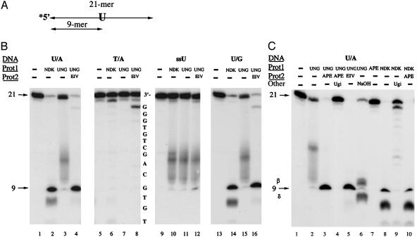
Fig. 1.
Uracil processing by eNDK. (A) Schematic view of the oligonucleotide substrate used in this experiment. Asterisk indicates the radioactively labeled terminus. (B) DNA cleavage analysis of U/A, T/A, ssU (single-stranded), and U/G oligonucleotides. Sequencing ladders prepared from the T/A substrate (partial sequence shown here) were run alongside samples. Enzymes and substrates used are indicated above lanes. The smeared appearance of UNG-treated DNA is because of instability of AP DNA and to the collapse of the helix from loss of base-stacking interactions. Reaction mixtures were separated on 20% sequencing gels, and the gels were subjected to autoradiography. (C) DNA cleavage analysis using the UDG inhibitor protein Ugi. See text for details.

