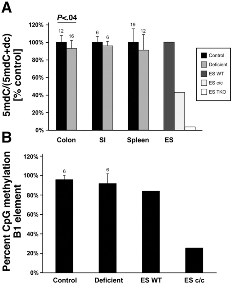
Figure 2.
Folate deficiency induces mild genomic hypomethylation of intestinal epithelial cells, but methylation levels of the B1 repetitive element are not significantly affected. (A) Genomic deoxymethylcytosine (5mdC) relative to total deoxycytosine (5mdC + dC) measured by mass spectrometry. To demonstrate relative changes, the average of the control group in each tissue was set as 100%. DNA from colon epithelial cells, small intestinal epithelial cells (SI), and spleen was analyzed. As controls we used wild-type ES cells (ES WT), ES cells knocked out for Dnmt1 (ES c/c), and ES cells deficient for Dnmt1, Dnmt3a, and Dnmt 3b (ES TKO). All folate-deficient tissues showed hypomethylation, and for colon epithelial cells this was statistically significant (Mann–Whitney U test), n above bars. (B) Pyrosequencing analysis of 4 CpG sites within the B1 repetitive element of colon DNA derived from control mice and folate-deficient mice (n = 6 each). DNA from ES WT and ES c/c were used as controls. Both groups contain 3 samples each from wild-type mice and Ung knockout mice. Shown is the average methylation level of all CpG sites combined; values are given as average ± SD. The folate-deficient samples show a small decrease in B1 methylation, which is not statistically significant (U test), n above bar.

