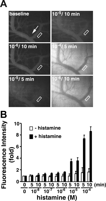
Fig. 1.
Histamine induces macromolecular leakage in cremaster venular microvessels of Sprague-Dawley (SD) rats. A: images recorded before (baseline) and 5 and 10 min after sequential application of increasing concentrations of histamine. White arrow indicates flow direction in veins. B: quantitation of changes in fluorescence intensity with (n = 11) or without (n = 2) histamine in extravascular space. Venular microvascular leakage was assessed by measurement of fluorescence intensity in area of interest (AOI; i.e., areas enclosed in rectangles in A). Values are means ± SE.

