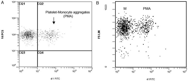
Fig. 3.
(A) Flow cytometric analysis of platelet-monocyte aggregates (PMAs) in whole blood displayed in a dual-fluorescence dot plot of CD14-PC5 and CD41-FITC gated on monocytes. Events in region CQ2 that are double positive are considered to be PMAs. Single-positive events are platelet-free monocytes. (B) Comparison of light scatter characteristics of CD14+CD41- and CD14+CD41+ populations. Population M represents platelet-free monocytes and PMA platelet-monocyte aggregates.

