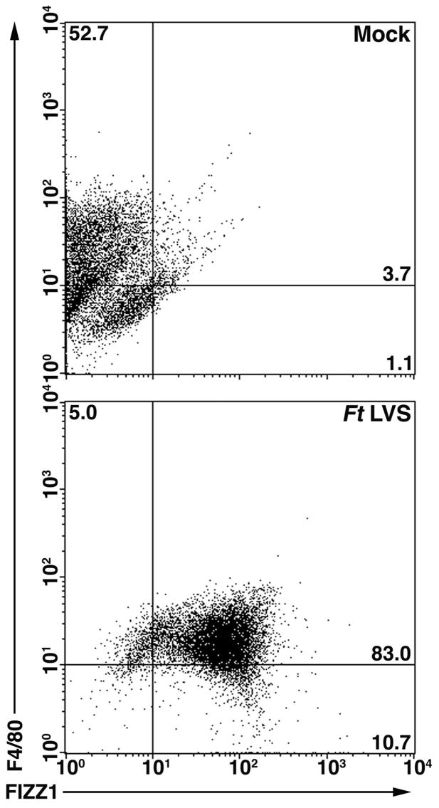
Figure 3.
In vivo Ft LVS infection results in AA-Mφ. C57BL/6 mice were administered saline or Ft LVS (10,000 CFU) i.p. Three days later, the mice were sacrificed and peritoneal macrophages were harvested and simultaneously stained for FIZZ1 and F4/80. Protein expression was determined by FACS analysis. The numbers in the quadrants indicate the percent of cells within that quadrant and have been rounded to the nearest one-tenth of a percent. Six mice were used for each treatment and data shown are from a single representative mouse.

