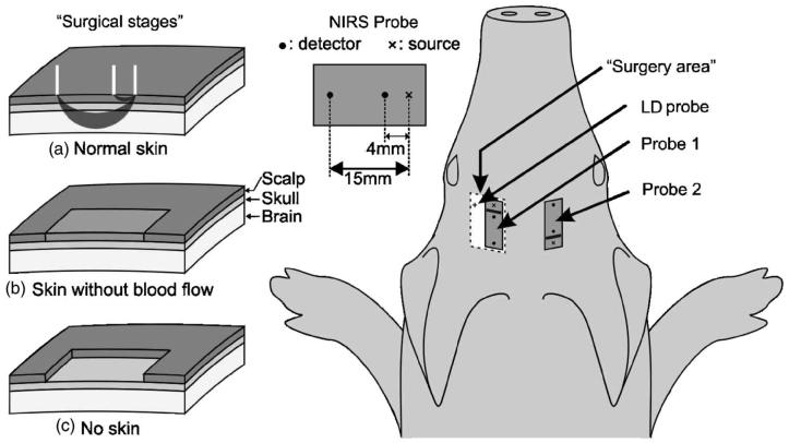
Fig. 1.
For protocol A, the two probes were positioned symmetrically on the two sides of the head. The surgery stages (a), (b) and (c) were performed only on the left hemisphere. The skin on the right side was not touched surgically, and probe 2 was not moved until the end of the measurements. For protocol B, we used only probe 1 and surgical stage (c). The Laser Doppler probe was positioned near the optical probe in a burr hole in the skull of the piglets.

