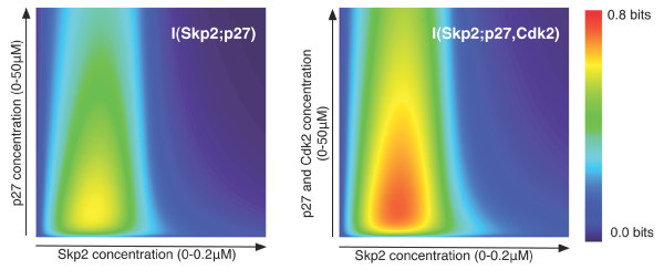
Figure 2.
Phase-space of cooperativity for the Cks1 adaptor protein. Both contour diagrams show the mutual information for different concentrations of Cks1, Skp2 and phosphorylated p27 (left panel) and for different concentrations of Cks1, Skp2, Cdk2 and phosphorylated p27 (right panel). In both panels, the concentration of Cks1 is kept fixed (0.1 μM) and the concentration of Skp2 and p27 vary between 0.0–0.2 μM and 0.0–50 μM respectively. In the right panel, Cdk2 varies together with p27, meaning that we assumed [p27] = [Cdk2] for all combinations. As can be observed, the signal between Skp2 and phosphorylated p27 is clearly constrained by the input concentrations. Moreover, when adding Cdk2, as shown in the right plot, the signal is reinforced.

