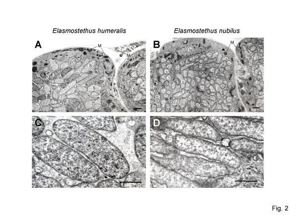
Figure 2.
Transmission electron microscopy of the symbiotic organ of acanthosomatid stinkbugs. (A) Midgut crypts of Elasmostethus humeralis. (B) Midgut crypts of Elasmostethus nubilus. (C) Symbiotic bacteria of E. humeralis. (D) Symbiotic bacteria of E. nubilus. Bars, 1 μm in (A) and (B); 0.3 μm in (C) and (D). Abbreviations: M, mitochondrion; N, nucleus; S, symbiont.

