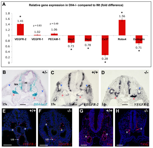
Figure 5.
Higher expression of VEGFR-2 and Robo-4 but lower expression of Endoglin in Dll4-/- embryos. (A) Relative quantitative gene expression in Dll4-/- embryos compared to Dll4+/+ (n = 4 per group) show increased expression of VEGFR-2 and Robo-4 and lower expression of Endoglin in the Dll4-/- mutants. Values were normalized to those of β-actin. (B-D) In situ hybridization on 9.0 dpc embryos cryosections show upregulation of VEGFR-2 specifically in Dll4-/- dorsal aortae (a). (B) In a thick section (50 μm) of a Dll4+/- embryo strong expression of Dll4 can be observed on the dorsal aortae and intersomitic vessels but no expression is detected on veins (v). (C) In a Dll4+/+ embryo VEGFR-2 is highly expressed in the veins and weakly expressed on the Dll4 expressing dorsal aortae. (D) In the absence of the arterial Dll4, VEGFR-2 is expressed at the same or at a higher level in the dorsal aortae compared to the anterior cardinal veins. (E, F) VEGFR1 (Flt1) in situ hybridization (in red) showing similar endothelial expression levels between control and mutant aortae (a) and umbilical arteries (ua). DAPI in blue. (G, H) Imunostaining for Endoglin (CD105) showing similar protein levels in endothelial cells of control and mutant embryos. Error bars represent st.dev. * indicates p < 0.05. (Scale Bars: 100 μm).

