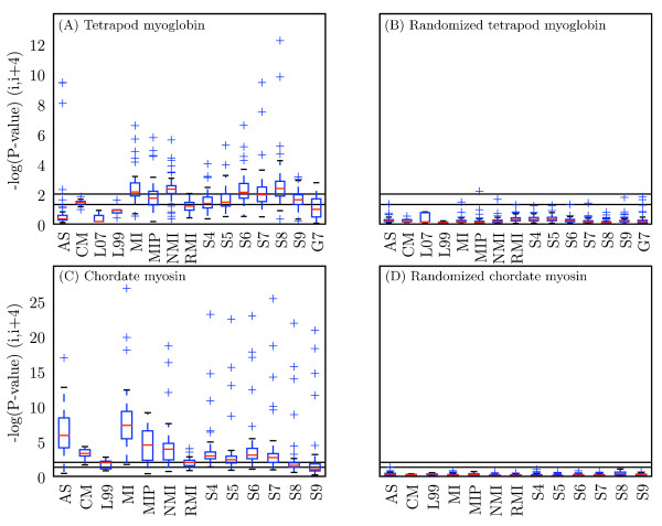
Figure 6.
Distribution of scores obtained with each method for all alphabets. Modified box plots illustrate the distribution of scores obtained with a given method over all alphabets. Values are -log(p-value) at (i, i + 4) for (A) tetrapod myoglobin, (B) randomized tetrapod myoglobin, (C) chordate myosin rod, and (D) randomized chordate myosin rod. Black lines indicate statistical significance threshold of α = 0.05 (bottom line) and α = 0.01 (top line). Red lines indicate the median value, and the top and bottom of the boxes indicate the upper and lower quartile values, respectively. Whiskers represent the largest and smallest values within 1.5 × IQR (inter-quartile range), and pluses represent outliers, or points outside of 1.5 × IQR. Methods with more condensed distributions are those that appear more robust to alphabet choice. AS: Ancestral States; L07: LnLCorr07; L99: LnLCorr99; MI: Mutual Information; NMI: Normalized Mutual Information; RMI: Resampled Mutual Information; Sn: Statistical Coupling Analysis, cutoff = n/10; MIP, Corrected Mutual Information; Gn: Generalized Continuous-Time Markov Process Coevolutionary Algorithm, ε = n/10; CM: CoMap.

