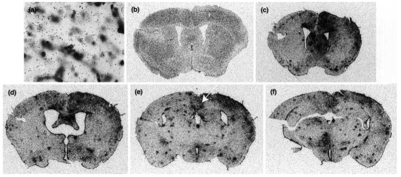
Fig. 4.
Induction of IRG2 expression reveals widespread activation caused by intracerebral injection of LPS/IFNγ. (a) Coronal section taken from a control mouse hybridized with a 35S-labeled sense TREM-2 riboprobe and counterstained as detailed in Fig. 3. in situ hybridization analysis using a 35S-labeled antisense IRG2 riboprobe was performed on coronal sections taken from the same unmanipulated control (b) as depicted in Figs 3(a) and (b) and 4(a), and on coronal sections taken from the same LPS/IFNγ-injected mice (c–f) as depicted in Figs 3(c) and (d). The focal plane is at the level of silver grains in the photographic emulsion. The white arrow in Fig. 4 shows the brain region depicted in Figs 3(c) and (d). Magnification × 40.

