
Figure 2.
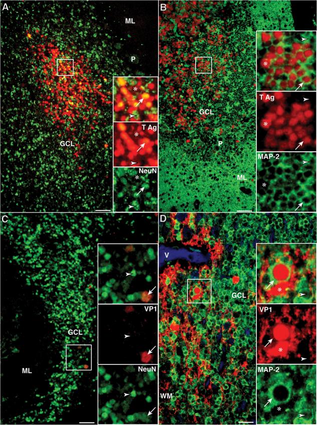
Presence of JCV T Ag and /or VP1 capsid protein in JCV-infected granule cell neurons (GCNs) of HIV+ PML patients (Bars: A–C = 50 μm; D = 25 μm). (A) Double IFA with rabbit polyclonal anti-SV40 T Ag (Alexa 568, red) and the neuronal marker mouse monoclonal anti-NeuN (Alexa 488, green) shows a cluster of hundreds of JCV-infected cells that express T Ag. Higher magnification views of cells in this cluster (square) shown in the upper inset, and viewed separately with the red (middle inset) and green channel (lower inset), show that most of the GCN, recognizable by their nuclear staining with NeuN, express T Ag in their nuclei (NeuN+/T Ag+ cells, arrows). In this cluster there are also a few JCV-infected glial cells, (NeuN−/T Ag+ cells, asterisk) and a few GCNs lacking T Ag (NeuN+/T Ag− cells, arrowhead). (B) The T Ag expressing GCNs can also be seen using double IFA with rabbit polyclonal anti-SV40 T Ag (Alexa 568, red) and a second neuronal marker mouse monoclonal anti-MAP-2 (Alexa 488, green). Because MAP-2 is mainly present in the cytoplasm, numerous MAP-2+/T Ag+ GCN are recognized by their red nuclei surrounded by a band of green cytoplasm (arrow, inset). Few MAP-2−/T Ag+ glial cells (red nuclei only, asterisk) and few non-infected GCNs (green cytoplasm only, arrowhead) are present. (C) Double IFA against JCV VP1 (SV40 antiserum, Alexa 568, red) and mouse monoclonal anti-NeuN (Alexa 488, green) reveals only isolated JCV-infected GCNs expressing VP1 capsid protein (NeuN+/VP1+, arrows), whereas the majority of GCNs do not express this protein (NeuN+/VP1−, arrowheads). (D) Similar results are seen with double IFA against VP1 (PAB597, Alexa 568, red) and rabbit polyclonal anti-MAP-2 (Alexa 488, green). Many MAP-2− glial cells displaying VP1 expression in their cytoplasm and nuclei (MAP-2−/VP1+, asterisk) are present in white mater (WM) with some extension in the granule cell layer (GCL). Rare GCNs (MAP-2+/VP1+, arrows) harboring nuclear expression of VP1 and cytoplasmic staining for MAP-2 can be found whereas most GCNs do not express VP1 (Map-2+/VP1−, arrowheads). (Asterisk, JCV-infected glial cell; Arrow, JCV-infected GCN; Arrowhead, GCN not expressing either T Ag or VP1. ML, molecular layer; P, Purkinje cells.)