
Figure 1. Chondrocytes express TRB3.
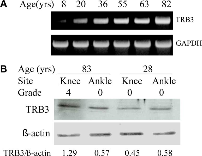
(A) Human chondrocytes isolated from donors of different ages were plated in monolayer culture. At confluency, RNA was isolated from these cells, RT-PCR was performed, and the cDNA products were stained with ethidium after separation on an agarose gel. GAPDH expression was used as a control. (B) Chondrocytes were isolated from matched pairs of knee and ankle cartilage from an old (83 years) and young (28 years) donor. Cell lysates were prepared after overnight incubation in serum-free media and were used for immunoblot analysis with anti-TRB3 antibody. Immunoblotting for actin was used as a protein loading control. Densitometry was used to quantitate the band intensity which is reported beneath the blots as a ratio of TRB3 to actin.