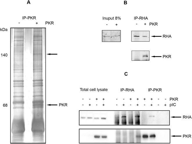
Figure 1. PKR associates with RHA.
(A) A silver-stained, SDS-PAGE gel of electrophoretically separated proteins immunoprecipitated with mouse monoclonal anti-PKR (IP-PKR) from pkr-null MEFs transformed with a human pkr construct either expressed (+) or silenced (−) and stimulated with pIC to activate the kinase. Arrows indicate the 68 kDa PKR and an associated 140 kDa protein. (B) A Western blot of proteins immunoprecipitated using a mouse monoclonal antibody to RHA (IP-RHA) from MEFs isolated form wild-type (+) or pkr-null (−) mice. Coimmunoprecipitated proteins are detected with rabbit polyclonal antibodies to RHA (upper panel), or murine PKR (lower panel). The levels of RHA expressed in the different cells were detected in whole cell lysates with the polyclonal anti-RHA antibody (Input). (C) A Western blot of proteins immunoprecipitated using antibodies to either PKR or RHA from pkr-null MEFs transformed with human PKR either expressed (+) or silenced (−) and differentially treated with pIC. Coimmunoprecipitated proteins are detected with opposing antibodies for RHA (upper panel) and PKR (lower panel).

