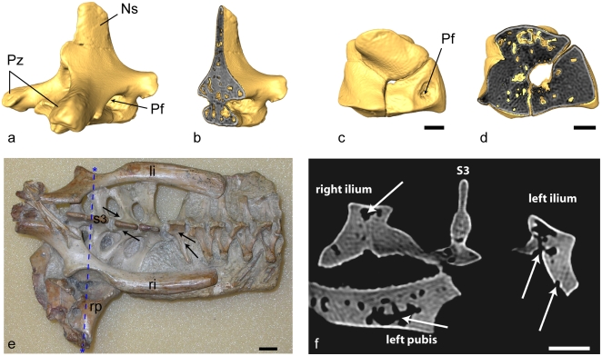
Figure 1. Micro-computed tomographic (CT) scans and photograph illustrating external pneumatic openings and typical pneumatic architecture in the ornithocheirid pterosaur Anhanguera santanae (AMNH 22555).
Vertebral (a, b), carpal (c, d), and pelvic (e, f) elements are characterized by the presence of thin cortical bone and large internal cavities (b, d, f). a, b, Mid-cervical (6th) vertebra in oblique craniolateral (a) and cutaway oblique craniolateral (b) views. Vertebral height = 5 cm. c, d, Left distal syncarpal in proximal (c) and cutaway proximal (d) views. e, dorsal view of block with pelvic elements, sacral vertebrae, and posterior dorsal vertebrae. Black arrows indicate the location of pneumatic foramina on select vertebrae (e) and white arrows indicate both pneumatic foramina and internal pneumatic cavities on pelvic elements (f). Asterisks on (e) delineate the location of the transverse section (dashed blue line) shown in (f). f, Transverse CT scan transect through pelvic block, showing pneumaticity of the sacral neural spine, ilia and left pubis. Note the large pneumatic opening on the surface of the left ilium. Scale bar (c–f) = 1 cm. li, left ilium; Ns, neural spine; Pf, pneumatic foramen; Pz, prezygapophysis; ri, right ilium; rp, right pubis; s3, sacral vertebra 3.

