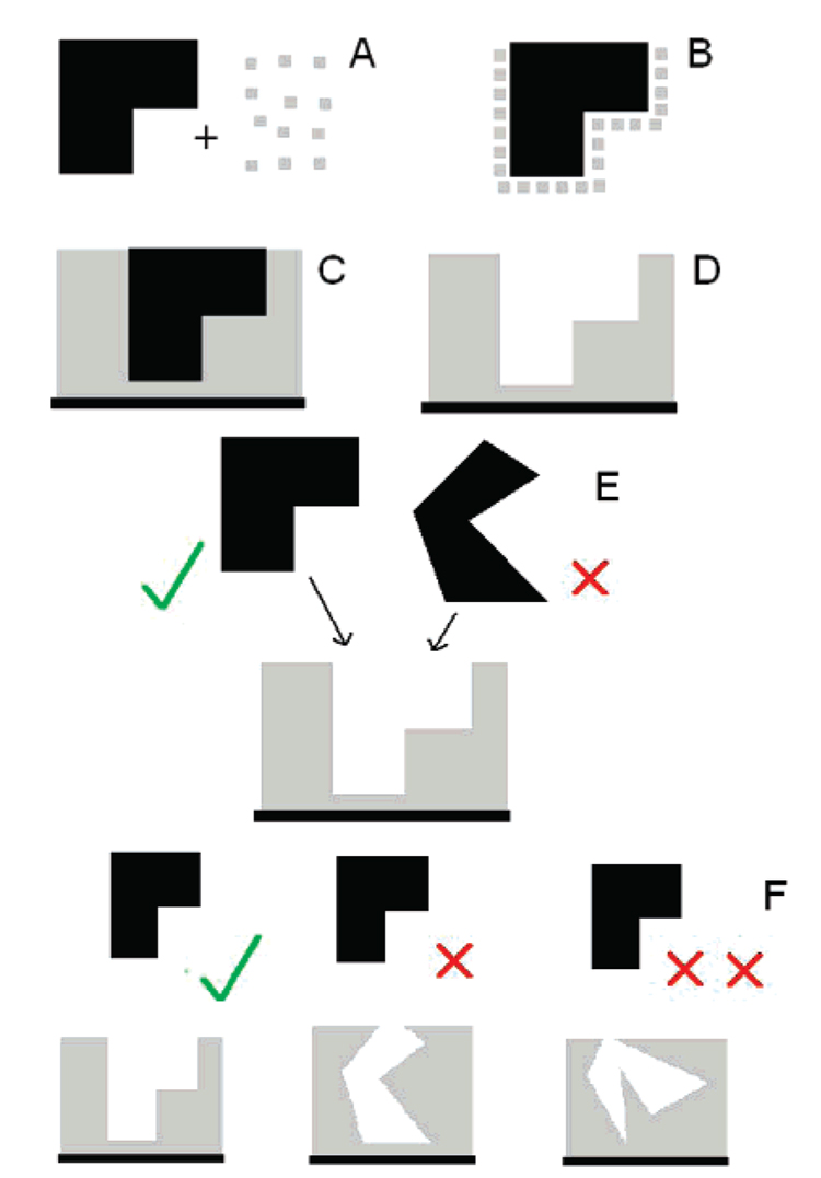
Figure 10.
Schematic representation of conformational protein molecular imprinting on a gold QCM crystal: (A) The template isoform of the protein (black) and APBA (gray) are mixed together to form a complex (B). Upon polymerization this complex is trapped within a poly(APBA) matrix which is adsorbed onto a surface (C). The template protein is extracted (D) to leave an imprint. When challenged with the template isoform or an alternative isoform of the same protein the imprint favors the template (E). The greater the difference between the protein structure used in the imprinting step and that used for rebinding, the less likely it is to bind (F).

