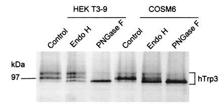
Figure 7.

Glycosylation and immunocytochemical analysis of Trp3 suggests the transmembrane topology shown in Fig. 3B3. HEKt3-9 cells expressing HA epitope-tagged hTrp3 in stable form or COS-M6 cells transiently expressing the same Trp, were metabolically labeled with [35S]Met/Cys. Cells were solubilized, and the HA-tagged proteins were immunoprecipitated by incubation with C12A5 monoclonal antibody and protein A-Sepharose, eluted with HA peptide, and either treated or not treated with Endo H or PN-glycosidase F as described in ref. 44. The resulting samples were analyzed by 9% SDS/PAGE, followed by autoradiography. The figure shows a digitally acquired picture of the autoradiogram processed with the aid of photofinish and powerpoint softwares and printed with a Canon CJ10 printer. Note that untreated [35S]hTrp3 runs as a complex set of bands that are converted to a single band of ≈97 kDa by treatment with PN glycosidase F, indicating that protein was glycosylated by the HEK cells.
