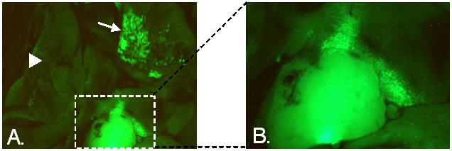
Figure 1.

A. Lungs of A/J female mouse 8 months after she had given birth following mating with a CAG/EGFP male, observed under fluorescent light. Fetally-derived GFP-expressing cells associated with a solid tumor are within the boxed area. Image of this tumor is enlarged in B. An apparent second lesion in a different lobe of the lung is indicated by the arrow. The arrowhead points to another lobe of the lung where fetally-derived cells are absent.
