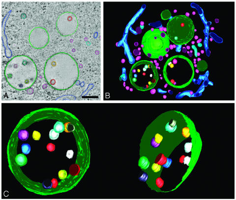
Fig. 2.
Three-dimensional model of MVB in RN cell. (A) Membranes in consecutive tomographic slices are manually traced to create a 3D model. Internal vesicles in the models are not perfectly round because of the manual tracing of the membranes. (Bar, 250 nm.) (B) Model view of the 3D reconstruction of the MVBs and surrounding area from Fig. 1. The space surrounding the MVBs is filled with tubules and vesicles. Movie 1 shows a movie of the 3D model. (C) Model view of the MVB tilted along two different angles. In the model on the right, the limiting membrane is partially removed to show the internal vesicles. The vesicles were each given a different color to trace them in each different view of the model.

