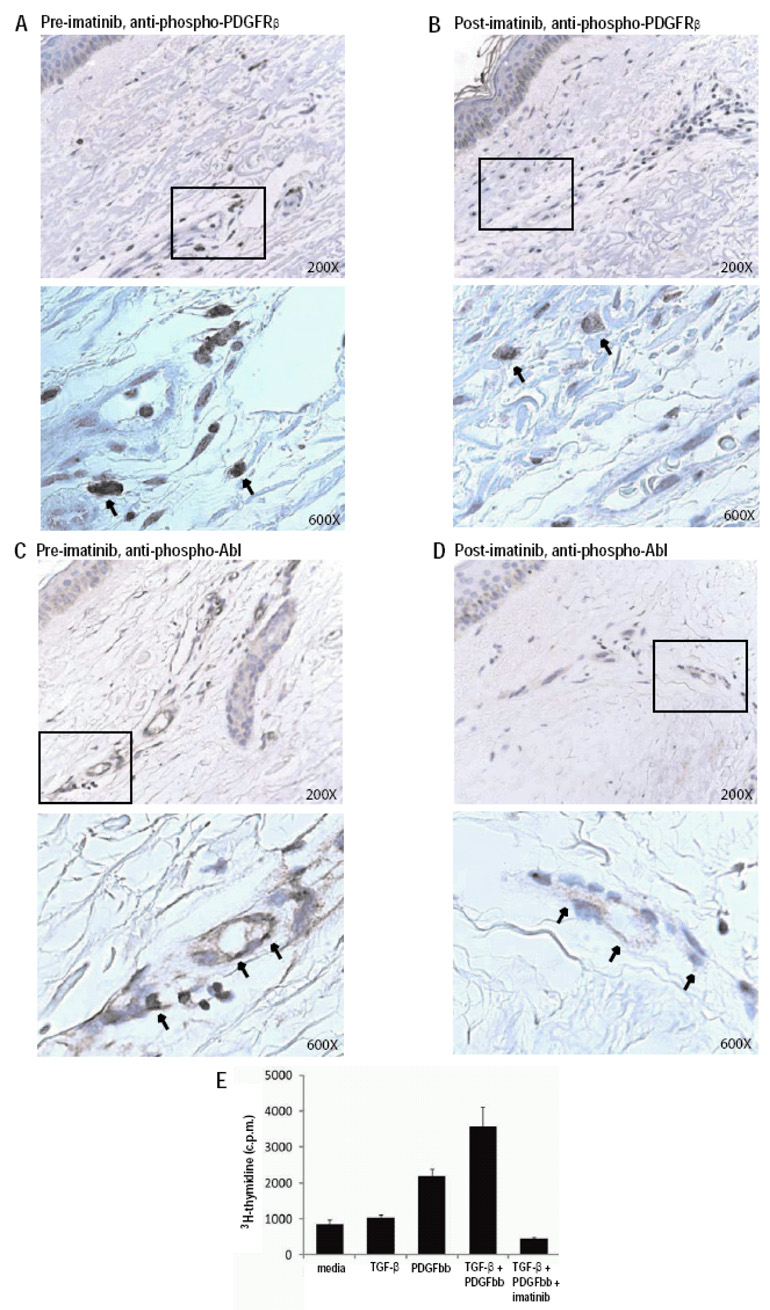
Figure 2. Imatinib reduces PDGFRβ and Abl activation in SSc skin and function in SSc fibroblasts.
(A–D) Immunohistochemical staining of serial skin biopsy samples obtained pretreatment (A,C) and one month following the initiation of imatinib treatment (B,D) with anti-phospho-PDGFRβ (A,B) and anti-phospho-Abl (C,D) antibodies. Boxed areas of upper panels (200× magnification), are presented at higher magnification in their corresponding lower panels (600×). Results are representative of those obtained from multiple sections from two independent patients. Phospho-PDGFRβ was observed in interstitial fibroblasts as well as perivascular spindle-like cells and some cells resembling mast cells. Phospho-Abl was observed in endothelial cells in small vessels and in scattered dermal fibroblasts. (E) Stimulation of a SSc fibroblast line with PDGF (10 ng/ml), TGF-β (0.5 ng/ml), PDGF + TGF-β, or PDGF + TGF-β+ imatinib (1 µM). Proliferation was quantitated after 48 hours by 3H-thymidine incorporation (Y axis). Results are representative of experiments performed on two independent SSc fibroblast lines, and similar results were obtained with normal fibroblast lines.

