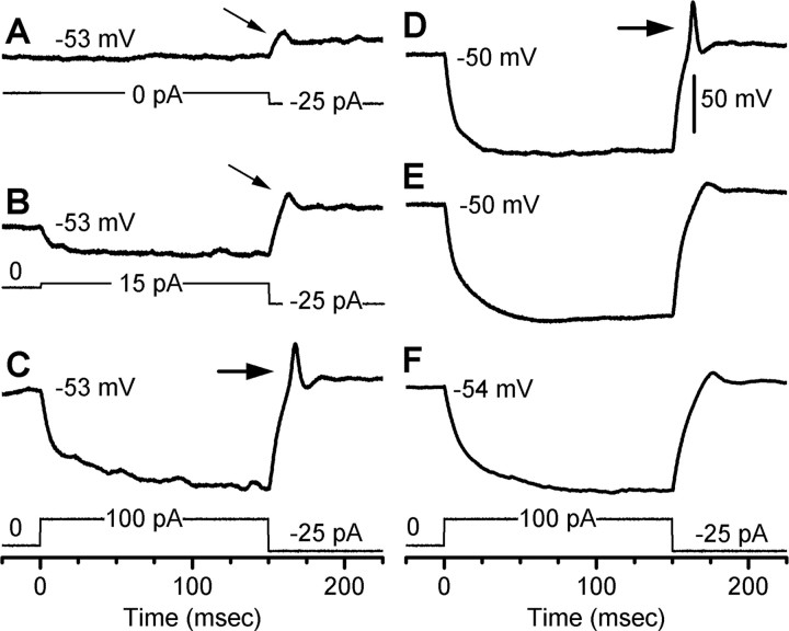
Figure 8.
Membrane potential spikes evoked by current steps in embryonic hair cells. Calibration applies to all traces. Resting potentials are indicated. A, Current-clamp response of an E16 cell stimulated with a -25 pA current step (protocol shown below). B, The same cell as in A was stimulated with a 15 pA prepulse to hyperpolarize the cell to -81 mV. Note the small passive depolarization with the step to -25 pA was similar to that of trace A (small arrows). C, The same cell as in A and B injected with a 100 pA prepulse to hyperpolarize the cell and relieve GNa inactivation. With the step to -25 pA, the cell evoked spike-like behavior (large arrow). D, Control trace from a different E16 cell. The protocol (below) was identical to that of C. E, The same cell and protocol as shown in D after bath exchange of sodium for NMDG+ to confirm the spike behavior was the result of GNa activation. F, A control trace from an E19 cell that lacked GNa and spike behavior.

