
Fig. 3.
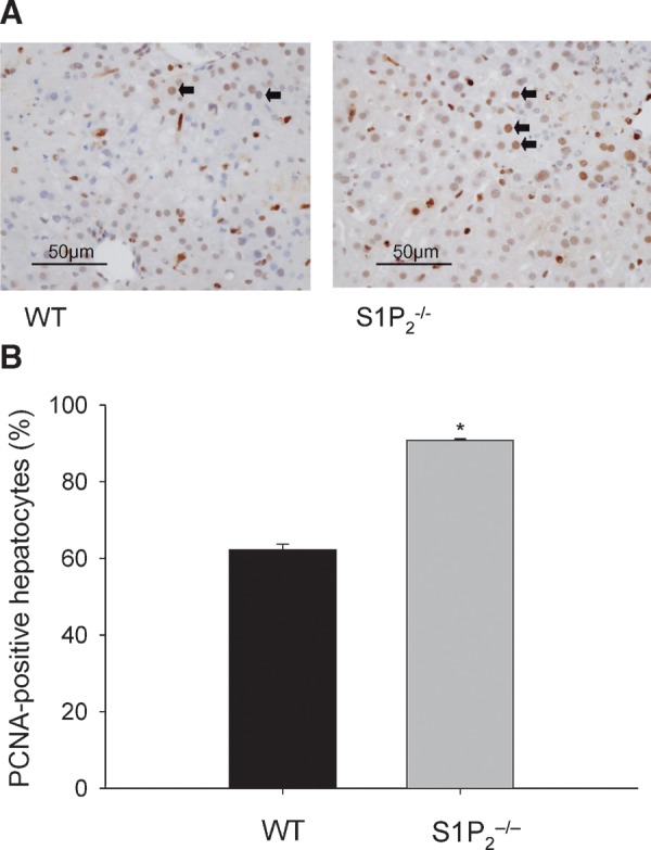
PCNA-positive hepatocytes of wild-type and S1P2−/− livers in acute liver injury induced by dimethylnitrosamine (DMN). A single injection of DMN was performed in wild-type (WT) and S1P2−/− mice. Immunohistochemical analysis of PCNA was done using a PCNA staining kit. Representative photomicrographs of the liver of wild-type and S1P2−/− mice at 48 h after DMN injection are shown (A). Bar = 50 μm. Arrows indicate positive hepatocytes. The percentage of PCNA-positive hepatocytes was determined with ten random areas at 400-fold magnification in each section (B). Columns and bars represent means ± SEM of four animals. The asterisk indicates a significant difference from wild type in Student's t-test (P < 0.05).