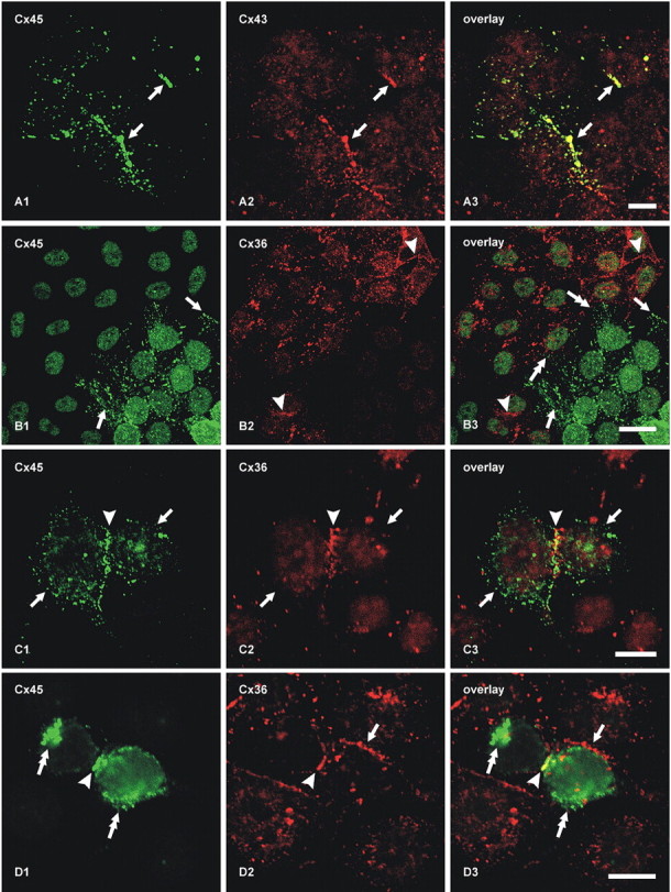
Figure 5.

Double-immunofluorescence labeling of connexins in cultures containing mixtures of HeLa cells expressing Cx45, Cx43, or Cx36. A, Coculture of cells stably expressing Cx45 with those expressing Cx43, showing colocalization of Cx45/Cx43 at abutments between Cx45-HeLa and Cx43-HeLa cells (A1–A3, arrows, yellow puncta in A3). B, Coculture of cells expressing Cx45 with those expressing Cx36, showing Cx45-positive puncta at cell appositions among colonies of Cx45-HeLa cells (B1, bottom right half of field and arrows) and Cx36-positive puncta at appositions among Cx36-HeLa cells (B2, top left half of field and arrowheads). There is a lack of Cx45/Cx36 colocalization at abutments between Cx45-HeLa and Cx36 HeLa cells (B3, double-headed arrows). C, HeLa cells stably expressing Cx36 and transiently transfected with Cx45, showing two apposed cells (arrows) expressing both connexins, and Cx45/Cx36 colocalization at points of cell–cell contact (arrowhead). D, Two HeLa cells stably expressing Cx36 and transiently transfected with Cx45, showing Cx45 localized intracellularly (D1, double-headed arrows), Cx36/Cx45 colocalization at points of cell–cell contact between cells expressing both connexins (arrowhead), and absence of Cx45 at points at which double-expressing cells make contacts with cells expressing only Cx36 (arrow). Scale bars, 10 μm.
