
Figure 8.
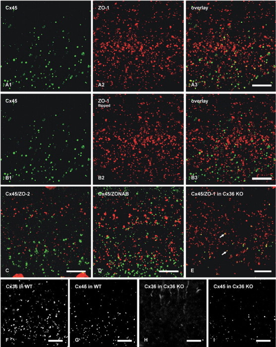
Laser-scanning confocal double immunofluorescence showing relationships of Cx45 with ZO-1, ZO-2, and ZONAB in the IPL of adult mouse retina, and Cx36, Cx45, and ZO-1 in the IPL of adult wild-type and Cx36 ko mice. Images show the IPL from inner (bottom) to outer (top) edge, and represent z-stacks of five confocal scans in A–D and single scans in E–I. Colocalization of green and red labeling is seen as yellow in image overlays. A, The same field (A1–A3) showing a high proportion of Cx45-positive puncta labeled for ZO-1. B, The same set of laser-scanning confocal double-immunofluorescence images as in A, showing minimal Cx45/ZO-1 colocalization in the IPL after horizontal flipping of the image showing labeling for ZO-1. C, Double-immunofluorescence overlay showing lack of Cx45 (green) colocalization with ZO-2 (red). D, Double-immunofluorescence overlay showing very few Cx45-positive puncta (green) labeled for ZONAB (red). E, Double-immunofluorescence overlay showing the persistence of Cx45/ZO-1 colocalization seen as yellow puncta (arrows) in the IPL of Cx36 ko retina. F–I, Confocal scans showing Cx36-puncta (F) and Cx45-puncta (G) in the IPL of wild-type retina and an absence of labeling for Cx36 (H) and a reduction of labeling for Cx45 (I) in IPL of Cx36 ko retina. Scale bars, 10 μm.