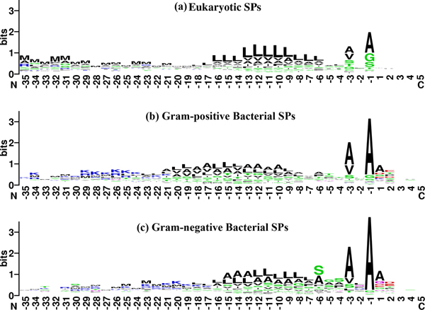
Figure 3.
Sequencelogos 37 of eukaryotic and bacterial (Gram-positive and Gram-negative) signal and mature peptides starting from posistion -35 to +5.[] The interface between position -1/+1 represents the SPase I cleavage site. The amino-acid residues are grouped and coloured based on the R group of their side-chain. Red denotes polar acidic amino-acid residues (D, E); Blue denotes polar basic amino-acid residues (K, R, H); Green denotes polar uncharged amino-acid residues (C, G, N, Q, S, T, Y); Black denotes non-polar hydrophobic amino-acid residues (A, F, I, L, M, P, V, W).

