
FIG. 1.
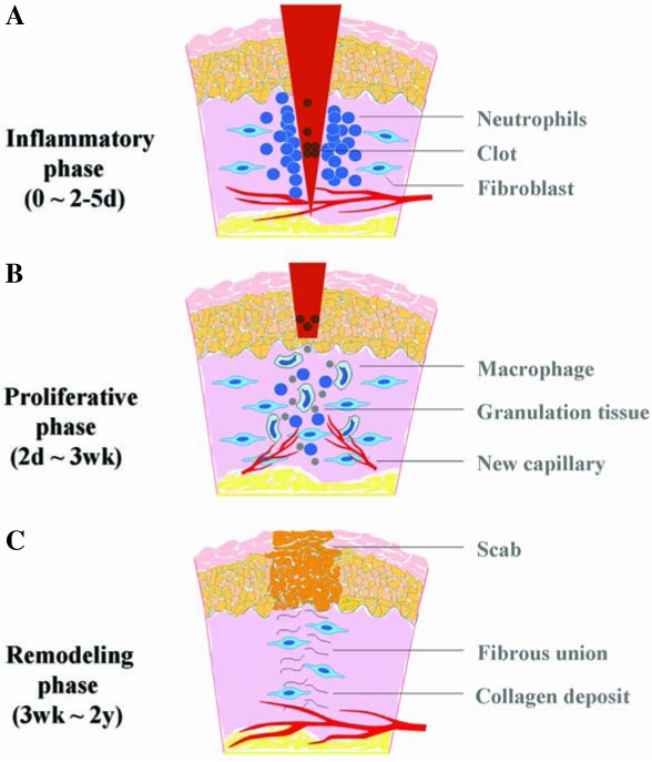
Schematic illustration of the three phases of acute wound healing. (A) Inflammatory phase: immediate to a few days. Immediately after the process of hemostasis, including vaso-constriction, platelet aggregation, and thromboplastin production, injured tissue undergoes vasodilation, and immune cells infiltrate and perform phagocytosis. (B) Proliferative phase: a few days to a few weeks. Fibroblasts lay a bed of collagen and fill the defect, while new capillaries grow (granulation). Injured tissue undergoes contraction. Wound edges pull together to reduce the defect. Afterward, epithelial cells migrate from the point of origin in all directions and cross the moist surface (epithelialization). (C) Remodeling phase: a few weeks to 2 years. During this phase, new collagen is formed and deposited, which increases tensile strength. (For interpretation of the references to color in this figure legend, the reader is referred to the web version of this article at www.liebertonline.com/ars).