Figure 1.

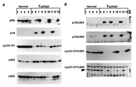
Expression and complex formation of p16/pRB pathway proteins in normal and tumor-derived breast epithelial cells. (A) Western blot analysis: exponentially growing normal and tumor cells were subjected to Western blot analysis using 50 μg of protein for each cell line in each lane of either a 6% (pRb), 13% (cyclin D1, cdk4, and cdk6), or 15% (p16) acrylamide gel and blotted as described. The same blot was reacted with cyclin D1, cdk4, and cdk6 affinity-purified antibodies. The blots were stripped between the three antibodies in 100 mM 2-mercaptoethanol, 62.5 mM Tris·HCl (pH 6.8), and 2% SDS for 30 min at 55°C. (B) Immune-complex formation: for immunoprecipitation followed by Western blot analysis, equal amounts of protein (500 μg) from cell lysates prepared from each cell line were immunoprecipitated with either monoclonal antibody to p16 (p16/cdk4 and p16/cdk6), polyclonal antibody to cyclin D1 (cyclin D1/cdk4), or a monoclonal antibody to cyclin D1 (cyclin D1/cdk6), coupled to protein A/G beads, and the immunoprecipitates were washed, boiled for 3 min, separated by SDS/13% PAGE, blotted to Immobilon membranes, and hybridized with either polyclonal antibody to cdk4 (p16/cdk4), polyclonal antibody to cdk6 (p16/cdk6 and cyclin D1/cdk6; arrow pointing to the complexed protein), or monoclonal antibody to cdk4 (cyclin D1/cdk4). The list of normal and tumor cell lines is presented in Table 1 using identical numbers.
