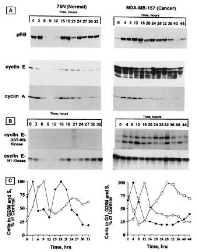
Figure 2.

Phosphorylation of pRb in synchronized population of tumor versus normal cells. Both cell types were synchronized by double thymidine block procedure. At the indicated times after release from double thymidine block, cell lysates were prepared and subjected to Western blot analysis (A) and histone H1 or GST-Rb kinase analysis (B). Protein (50 μg) for each time point was applied to each lane of either a 6% (pRb) or 10% (cyclins E and A) acrylamide gel and blotted as described. The same blot was reacted with cyclin E monoclonal (HE12) and cyclin A affinity-purified polyclonal antibodies. The blots were stripped between the two assays as described for Fig. 1. For kinase activity, equal amounts of proteins (600 μg) from cell lysates prepared from each cell line at the indicated times were immunoprecipitated with anti-cyclin E (polyclonal) coupled to protein A beads using either histone H1 or purified GST-Rb as substrates. (C) The relative percentage of cells in different phases of the cell cycle for each cell line at various times after release from double thymidine block was calculated from flow cytometric measurements of DNA content. ♦, cells in S phase; ○, cells in G2/M phase; □, cells in G1 phase.
