Fig. 5.
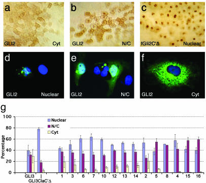
Subcellular localization of GLI2 protein in animal cap cells of injected gastrula (stage 11) frog embryos. The normal and variant proteins were detected by using an antibody against the N-terminal myc tag. Representative examples of blastomere staining patterns are shown and quantified. Background levels were essentially none. (a) Prominent cytoplasmic staining, compared with combined nuclear and cytoplasmic staining (b). As a control for primarily nuclear staining (c) we used a frog Gli2 construct deleted for its C terminus (fGli2C′Δ) and previously shown to exhibit nuclear accumulation (6). A similar subcellular localization in transfected COS-7 cells was scored as primarily nuclear (d), both nuclear and cytoplasmic (e), or exclusively cytoplasmic (f). (g) Histograms showing the distribution of the different alleles in transfected COS-7 cells. Each allele was transfected three or four times, and >100 cells were counted in each experiment.

