Fig. 6.
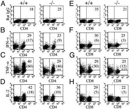
Reduced levels of TNF-α in activated IFN-β-/- T cells. Splenocytes were stimulated with mouse recombinant anti-CD3 for 48 h, followed by PMA and ionomycin for 4 h. Cells were then simultaneously labeled with cell-surface Abs for CD4 (A-D) or CD8 (E-H) and stained for either a nonspecific antibody with the same isotype (A and E) or intracellular cytokine antibodies as indicated. Values shown are the lymphocyte population as a percentage of total splenic population. Numbers in parentheses represent the percentage of either CD4- or CD8-positive cytokine-secreting cells. Statistical analysis was by Student's t test; **, P = 0.008; ***, P = 0.001. +/+, IFN-β+/+; -/-, IFN-β-/-. Data are representative from IFN-β+/+ (n = 5) and IFN-β-/- (n = 5) mice.

