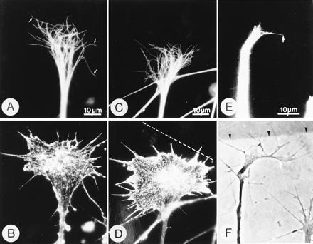
Figure 1.

Microtubules become reoriented in growth cones at tenascin borders. Phase-contrast and immunofluorescence micrographs of growth cones immunostained with mAb YL 1/2, which stains microtubules (A, C, and E) and mAb N350, which stains actin (B and D). (F) Phase-contrast image of the living growth cone shown in E. In growth cones growing on laminin (A and B), microtubules are splayed out in the central domain and extend, individually, far into the peripheral domain and occasionally insert into filopodia (arrows in A). In this growth cone, 52% of the microtubules had terminations in the left-hand area of the growth cone and therefore, by the definition given in Materials and Methods, the distribution is symmetrical. However, in growth cones at laminin-tenascin borders, microtubules become asymmetrically distributed (C and E). In C and D, the actin staining (D) reveals the growth cone morphology which suggests that the axon approached the border at a slight angle and that the right hand side of the growth cone has contacted the border, indicated by the broken line. The microtubules in this growth cone (C) are asymmetrically distributed (65% of the microtubules terminate in the right-hand half). In micrographs E and F the growth cone has clearly turned to the right and the microtubule asymmetry is obvious (arrow in E). Arrows in E and F are at the same location. The laminin–tenascin border is visualized with colloidal gold (arrowheads).
