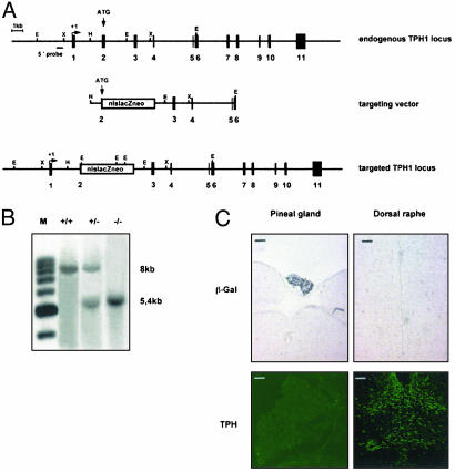
Fig. 1.
Targeted disruption of the tph1 gene. (A) Strategy for targeting the tph gene. (Top) The endogenous tph1 locus. (Middle) The targeting construct. (Bottom) The structure after homologous recombination. (B) Southern blot analysis of the wild-type (+/+), heterozygous (+/-), and homozygous (-/-) alleles. The 5′ probe, as indicated in A, recognizes a sequence outside the recombined region. An 8-kb EcoRI fragment is detected in the wild-type allele, and a 5.4-kb fragment is detected in the recombined allele. (C) Immunohistochemical analysis of the pineal gland and the dorsal raphe nucleus of homozygous tph1 mutants. (Upper) β-Gal immunoreactivity. (Lower) TPH immunoreactivity as described (39). (Scale bars = 0.1 mm.)

