
Figure 7.
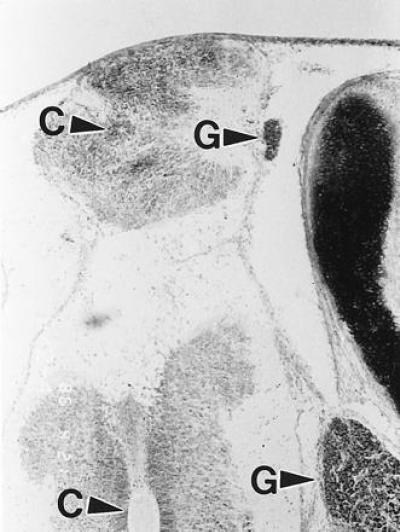
A 9-day embryo treated with HcysTD showed a spina bifida grossly similar to that shown in Fig. 5. Histologic sections of this lesion stained with the Spicer method showed a small, abnormal duplication of a fairly normal cord; the redundant branch was separated from the outside by a thin membrane. C, central canal; G, spinal ganglion.